

How Does Amoeba Get Energy
What is an amoeba?
"Amoeba" is a term that describes a elementary eukaryotic organism that moves in a characteristic crawling fashion. However, a comparison of the genetic content of the various amoebas shows that these organisms are not necessarily closely related to each other.
What does an amoeba look similar?
All living organisms tin can exist broadly divided into two groups — prokaryotes and eukaryotes — which are distinguished past the relative complexity of their cells. Eukaryotes are highly organized unicellular or multicellular organisms, such as animals and plants. Prokaryotes, on the other hand, are basic unmarried-celled organisms, such as bacteria and archaea.
Related: Foreign single-celled life-course has a truly bizarre genome
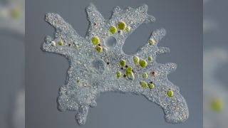
Amoebas are eukaryotes. Their single cells, similar those of other eukaryotes, possess certain feature features: Their cellular contents are enclosed within a cell membrane, and their DNA is packaged into a central cellular compartment chosen the nucleus, according to a 2014 report published in the journal BMC Biology (opens in new tab). In addition, they contain specialized structures called organelles, which perform a range of cellular functions including free energy production and protein transport.
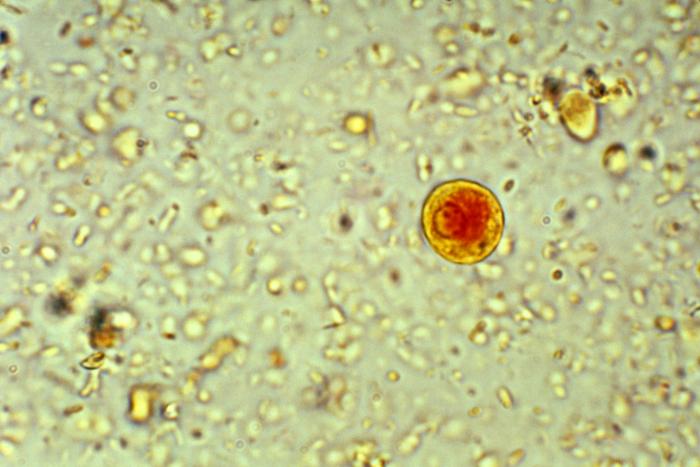
Virtually of these organelles are common to all eukaryotic cells, merely there are a few exceptions. For case, the parasitic amoebas Entamoeba histolytica, which crusade amoebic dysentery in humans, do not have the golgi apparatus, the organelle responsible for modifying and transporting proteins, according to a 2005 written report published in The Periodical of Biological Chemistry (opens in new tab). Researchers found that Due east. histolytica instead contain golgi-like compartments or vesicles — small-scale fluid-filled pouches — that execute similar functions.
There are also amoebas that don't have mitochondria, the organelle responsible for generating cellular energy, because they live in environments lacking in oxygen, or "anoxic conditions," Sutherland Maciver, a reader in the department of biomedical sciences at the University of Edinburgh, told Live Science.
According to a 2014 review published in the journal Biochemie (opens in new tab), these organisms without mitochondria tin comprise organelles called hydrogenosomes or mitosomes, which are related to mitochondria simply are thought to be highly altered versions of the organelle. This is the case for Due east. histolytica and the free-living amoeba Mastigamoeba balamuthi, which doesn't depend on other organisms for survival.
How does an amoeba motility?
Structurally, amoebas closely resemble the cells of college organisms. "They are similar our cells, and in fact, when they are moving they look very much like our white claret cells," Maciver said. (White blood cells are immune cells that help defend the body against disease.)
Like our white blood cells, amoebas move using pseudopodia, which translates to "false anxiety" in Latin. These brusk-lived, outward projections of the cytoplasm — the semifluid material inside the cell membrane — help amoebas to grip a surface and propel themselves forward. As the pseudopodium moves out along a surface in 1 direction, the back stop of the amoeba contracts, Maciver said.
"As it contracts, information technology does ii things," he said. "The contraction pushes the cytoplasm frontward to fill the expanding pseudopod, but the contraction besides pulls upwards adhesions at the back end of the cell." Maciver describes these adhesions between an amoeba and the surface on which information technology moves as concrete molecular adhesions, which are constantly formed at the front and broken at the back. This movement, using pseudopodia, is a characteristic that unites diverse amoebas and distinguishes them from other protists — simple eukaryotic organisms similar amoebas that are not plants, animals or fungi.
There are four unlike types of pseudopodia seen among amoebas: filopodia, lobopodia, rhizopodia and axopodia, according to Human Parasitology (opens in new tab) (Academic Printing/Elsevier, 2019). The most mutual course of parasitic amoebas have lobopodia, which are wide, blunt cytoplasmic projections, while filopodia are thin, thread-like projections.
Rhizopodia, also known every bit reticulopodia, are thin filament-like projections that mesh together, and axopodia are rigid and strengthened by an array of microtubular structures called axonemes, according to Ecology and Nomenclature of Northern American Freshwater Invertebrates (opens in new tab) (Academic Press, 2001). Other pseudopods are supported by structural tube-shaped elements known equally microtubules, which are responsible for executing cell movements.
Related: Robert Hooke: English scientist who discovered the cell
Amoebas can as well use their pseudopodia to feed. A 1995 study, published in the journal Applied and Ecology Microbiology (opens in new tab), gives the example of the soil-domicile amoeba Acanthamoeba castellanii, which ingests both solids and liquids using its pseudopodia. The process of ingesting solid fabric is called phagocytosis, and the process of engulfing drops of liquid is known as pinocytosis, also known as prison cell drinking, according to Dosage Form Design Considerations (opens in new tab) (Bookish Press/Elsevier, 2018).
"Nigh of the known amoebae eat bacteria," Maciver told Live Science. He explained that amoebas have receptors on their prison cell surface that demark to bacteria, which are then taken into amoebas by phagocytosis, usually at the rear of the cell.
In the case of giant amoebas, such as Amoeba proteus, the process of phagocytosis is slightly different, co-ordinate to Maciver. Giant amoebae engulf their casualty "by the willful gathering of pseudopods effectually the bacteria." In both cases, equally the bacteria is drawn in, the cell membrane that surrounds it pinches off to form an intra-cellular compartment chosen the vacuole.
How are amoebas classified?
For centuries, the diverse systems of classifying organisms, including amoebas, were based on similarities in appreciable characteristics and morphology. "There isn't actually a coherent group of organisms called the amoebae," Maciver said. "Rather, amoebae are any protozoan cells that motion past itch." (The term "protozoa" refers to a subset of protists, which over again are uncomplicated eukaryotic organisms that are not plants, animals or fungi, Live Science previously reported.)
Historically, amoebas were classified together in a single taxonomic group called Sarcodina, distinguished by their apply of pseudopodia. Sarcodina amoebas were and so subdivided based on the specific blazon of pseudopodia they used, according to a 2008 commodity published in the journal Protistology (opens in new tab). However, this system of nomenclature didn't capture the evolutionary relationships betwixt the diverse amoebas — it was not a family tree, so to speak.
Molecular phylogenetics changed the course of taxonomic classification for eukaryotes. By comparing the similarities and differences in particular Dna sequences within organisms, scientists were able discern how closely related they were, co-ordinate to a 2020 review in the journal Trends in Ecology & Evolution (opens in new tab).
Early on analyses compared the DNA sequences that encode part of the ribosome, the site of poly peptide synthesis in a cell; specifically, scientists looked at the genes for the and then-called 18S subunit of ribosomes, or "SSU rDNA." Based on the analyses of SSU rDNA and other DNA sequences, eukaryotic organisms are now organized in a mode that meliorate represents their evolutionary relationships — the phylogenetic tree, according to the 2008 Protistology article.
Each lineage in a phylogenetic tree is depicted past a branched structure. In this system, the offset levels are known equally "supergroups." Fabien Burki, writer of a 2014 review commodity published in the journal Cold Spring Harbor Perspectives in Biology, (opens in new tab) described these supergroups as the "building blocks" of the tree.
Burki listed five supergroups for eukaryotic organisms: Ophiskontha, Amoebozoa, Excavata, Archaeplastida and SAR, which includes iii subgroups named Stramenopiles, Alveolata and Rhizaria. Animals and fungi are in the group Ophiskontha. Amoeboid protists and some parasitic lineages that lack mitochondria are part of Amoebozoa. Together, Ophiskontha and Amoebozoa course a larger supergroup called Amorphea, co-ordinate to the review in the periodical Trends in Ecology & Evolution.
Heterotrophic protists — organisms that take in nutrients from other organisms — are part of Excavata, while plants and nearly other photosynthetic organisms are part of Archaeplastida, according to The Encyclopedia of Evolutionary Biology (opens in new tab) (Bookish Press/Elsevier, 2016).
"If you look at the great diversity of the protists, you can run across that at that place are amoebae in near all the groups," Maciver said. "There's even an amoeboid organism within the chocolate-brown algae [Labyrinthula]." Nearly amoebas are nowadays within the Amoebozoa group, though, Maciver said. In add-on, he noted that amoebas are likewise nowadays within Rhizaria and Excavata. Nucleariids, a group of amoebae with filopodia, belong to the Opisthokonta supergroup, for instance, and Labyrinthulids fit inside the Stramenopiles.
Why are amoebas important?
Amoebas are known to cause a range of human diseases. Amebiasis, or amoebic dysentery, is an infection acquired by E. histolytica, a human intestinal parasite, according to the Centers for Affliction Control and Prevention (opens in new tab) (CDC). According to the medical database StatPearls (opens in new tab), E. histolytica can invade the colon wall and cause colitis, where the inner lining of the colon becomes inflamed, and the parasite tin can crusade severe diarrhea and dysentery.
Though East. histolytica infections can occur anywhere in the world, it is most common in tropical regions that have substandard sanitation systems and crowded conditions.
Contact lens wearers are potentially at risk of a rare infection of the cornea called Acanthamoeba keratitis. According to the CDC (opens in new tab), species in the Acanthamoeba genus are free-living and are commonly establish in soil, air and h2o. Poor contact lens hygiene practices, such as improper storage, handling and disinfection or swimming with lenses, are some of the risk factors for the disease, the CDC states. Contact lens users can reduce their take a chance of infection by wearing and cleaning their lenses as prescribed past their center care provider, and removing their lenses before any activity involving contact with water, including showering, using a hot tub or swimming.
While the initial symptoms include redness, itchiness and blurred vision, if left untreated the infection can cause severe hurting and lead to vision loss, co-ordinate to the CDC.
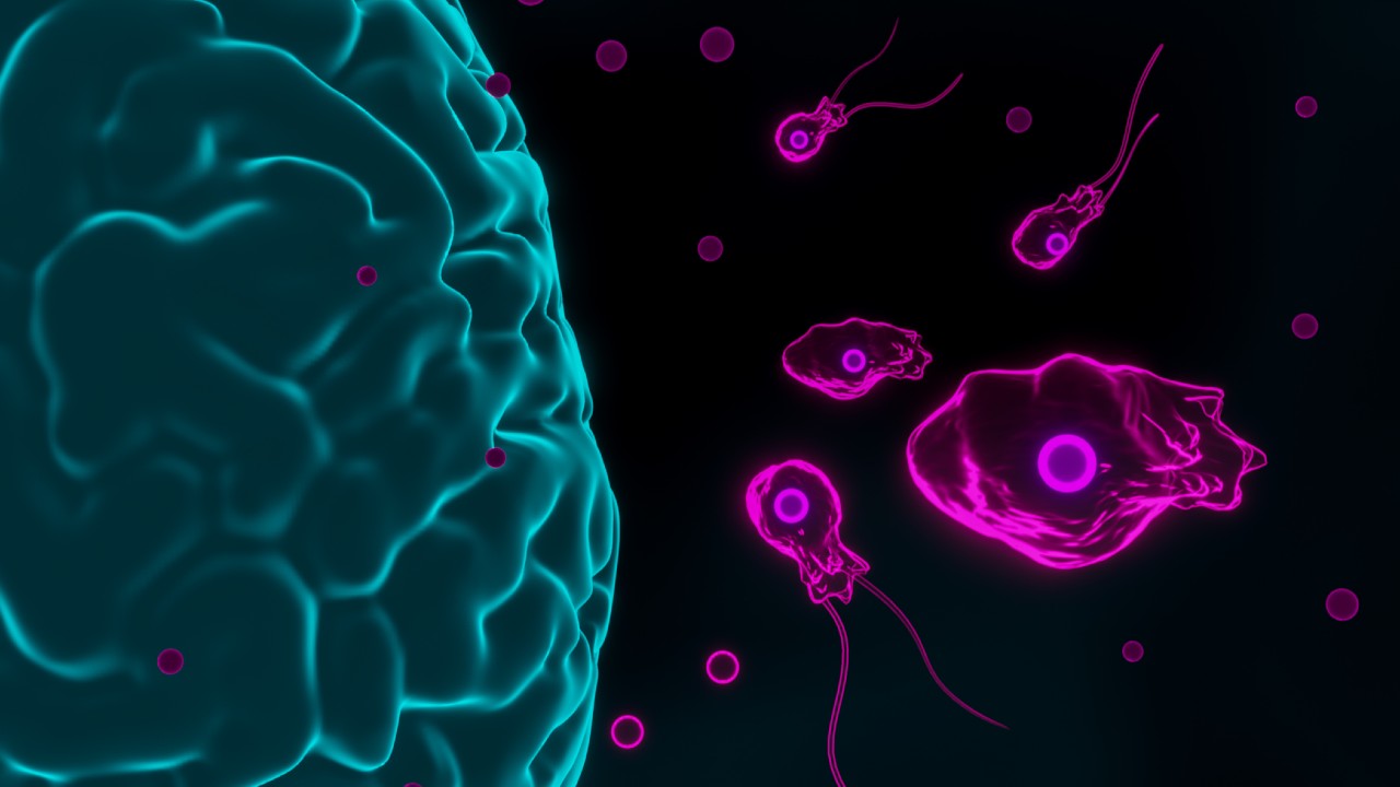
Amoebas also cause unlike infections of the brain. Naegleria fowleri , which has been dubbed "the brain-eating amoeba," causes primary amoebic meningoencephalitis (PAM). Though the disease is rare, information technology is almost always fatal, according to the CDC (opens in new tab). Early on symptoms include fever and vomiting, and the illness ultimately progresses to more severe symptoms such as hallucinations and coma. North. fowleri is present in warm freshwater bodies such as hot springs, lakes and rivers, or in poorly chlorinated swimming pools or contaminated, hot tap water. These amoebas enter from the olfactory organ and travel to the brain. Even so, the infection can't be contracted past swallowing water, co-ordinate to the CDC.
Some other amoeba, Balamuthia mandrillaris, tin can cause a brain infection known as granulomatous amoebic encephalitis (GAE). Balamuthia infections are rare only are frequently fatal. Electric current estimates advise that the infection has a death rate of xc%, the CDC states (opens in new tab).
Early symptoms include headaches, nausea and low-grade fever, partial paralysis, seizures and speech difficulties. B. mandrillaris is plant in the soil and can enter the torso through open wounds or when people breathe in contaminated grit, co-ordinate to the CDC. Since the amoeba was discovered in the 1980s, about 200 cases of the infection have been reported worldwide; this includes more than 100 confirmed cases in the U.S.
Amoebas can also play host to leaner that are pathogenic to humans, and assist those bacteria spread. Bacterial pathogens, such equally Legionella, which can cause pneumonia- and flu-like illnesses, can resist digestion when consumed by amoebas, according to a 2018 written report in the journal Front Cell Infection Microbiology (opens in new tab). Instead, the bacteria are released intact from vacuoles into an amoeba'due south cytoplasm, where they proliferate inside the cell. In such cases, bacteria tin get resistant to treatments designed to control their numbers, including chlorine treatment of water.
Maciver cites the example of cooling towers as a place where both amoebas and these bacteria can grow. Cooling towers tend to expel water droplets, which passersby tin breathe in. "What'south known to happen on many occasions, is that we breathe in a droplet of water containing an amoeba that is full of these pathogens [Legionella]," he said. If bacteria enter the torso of an immunocompromised individual in such a manner, they can ultimately infect macrophages, i of the allowed system's many defensive cells.
"A macrophage not only looks similar an amoeba, its biochemical pathways and jail cell biological science are quite similar," Maciver said. "So the aforementioned programmed events that allow the bacteria to escape the amoeba at present operate to allow Legionella to escape the macrophage."
Apart from their roles in human being disease, amoebas are as well an important part of the soil ecosystem. Amoebas casualty on harmful leaner and regulate their population in the soil, according to a 2021 review in the journal Practical and Environmental Microbiology (opens in new tab).
Amoebas are also important for recycling nutrients in the soil. According to Maciver, when nutrients go available they are taken up by bacteria, which "effectively lock upwardly all the nutrients in bacterial mass." When bacteria are consumed, nutrients are released back into the soil. "If y'all have a cycle whereby amoeba eat bacteria, the overall effect is to increase nutrient availability for plants," Maciver said.
Additional resources and readings
- The Amoeba in the Room: Lives of the Microbes (opens in new tab) explores the incredible diversity of the microbial earth.
- Learn more about different species of protist in Protists: Algae, Amoebas, Plankton, and Other Protists (A Form of Their Own). (opens in new tab)
- Read about the many microbes that live inside our bodies in Ed Yong's I Comprise Multitudes: The Microbes Within Us and a Grander View of Life (opens in new tab).
Bibliography
Acharya, P. C., Fernandes, C., Mallik, South., Mishra, B., & Tekade, R. Thousand. (2018). Physiologic Factors Related to Drug Absorption. In Dosage form blueprint considerations (Vol. i, pp. 117–147). chapter, Bookish Press/Elsevier.
Avery, S. V., Harwood, J. 50., & Lloyd, D. (1995). Quantification and label of phagocytosis in the soil amoeba Acanthamoeba castellanii past flow cytometry. Applied and Environmental Microbiology, 61(3), 1124–1132. https://doi.org/https://world wide web.ncbi.nlm.nih.gov/pmc/manufactures/PMC1388394/pdf/hw1124.pdf (opens in new tab)
Baum, D. A., & Baum, B. (2014). An inside-out origin for the eukaryotic cell. BMC Biology, 12(1). https://doi.org/ten.1186/s12915-014-0076-2 (opens in new tab)
Bogitsh, B. J., Carter, C. E., & Oeltmann, T. N. (2019). General Characteristics of the Euprotista (Protozoa). In Human parasitology (fifth edition) (5th ed.). chapter, Academic Press.
Bredeston, 50. Thousand., Caffaro, C. E., Samuelson, J., & Hirschberg, C. B. (2005). Golgi and endoplasmic reticulum functions take place in different subcellular compartments of Entamoeba histolytica. Journal of Biological Chemical science, 280(37), 32168–32176. https://doi.org/x.1074/jbc.m507035200 (opens in new tab)
Burki, F. (2014). The Eukaryotic Tree of life from a global phylogenomic perspective. Common cold Spring Harbor Perspectives in Biology, six(five). https://doi.org/10.1101/cshperspect.a016147 (opens in new tab)
Burki, F., Roger, A. J., Chocolate-brown, Thousand. Westward., & Simpson, A. G. B. (2019). The New Tree of Eukaryotes. Trends in Environmental & Evolution, 35(1), 43–55. https://doi.org/10.1016/j.tree.2019.08.008 (opens in new tab)
Centers for Affliction Control and Prevention. (2010, November 2). Acanthamoeba keratitis FAQs. Centers for Disease Control and Prevention. Retrieved Jan 28, 2022, from https://www.cdc.gov/parasites/acanthamoeba/gen_info/acanthamoeba_keratitis.html (opens in new tab)
Centers for Disease Command and Prevention. (2020, June 2). Balamuthia mandrillaris - Illness & symptoms. Centers for Disease Control and Prevention. Retrieved January 28, 2022, from https://www.cdc.gov/parasites/balamuthia/affliction.html (opens in new tab)
Centers for Disease Command and Prevention. (2021, December 3). Parasites - amebiasis. Centers for Disease Control and Prevention. Retrieved January 28, 2022, from https://www.cdc.gov/parasites/amebiasis/index.html (opens in new tab)
Chou, A., & Austin, R. L. (2021, April 25). Entamoeba histolytica. StatPearls [Internet]. Retrieved Jan 28, 2022, from https://www.ncbi.nlm.nih.gov/books/NBK557718/ (opens in new tab)
Makiuchi, T., & Nozaki, T. (2014). Highly divergent mitochondrion-related organelles in anaerobic parasitic protozoa. Biochimie, 100, 3–17. https://doi.org/10.1016/j.biochi.2013.11.018 (opens in new tab)
McCourt, R. (2016). Archaeplastida: Diversification of Red Algae and the Green Plant Lineage. In Encyclopedia of evolutionary biology (pp. 101–106). entry, Elsevier, Academic Printing. https://world wide web.sciencedirect.com/scientific discipline/article/pii/B9780128000496002547 (opens in new tab)
Oliva, G., Sahr, T., & Buchrieser, C. (2018). The life cycle of L. Pneumophila: Cellular differentiation is linked to virulence and metabolism. Frontiers in Cellular and Infection Microbiology, viii. https://doi.org/ten.3389/fcimb.2018.00003 (opens in new tab)
Pawlowski, J. (2008). The twilight of Sarcodina: a molecular perspective on the polyphyletic origin of amoeboid protists. Protistology, 281–302. https://doi.org/http://www.zin.ru/journals/protistology/num5_4/pawlowski.pdf (opens in new tab)
Shi, Y., Queller, D. C., Tian, Y., Zhang, South., Yan, Q., He, Z., He, Z., Wu, C., Wang, C., & Shu, 50. (2021). The ecology and evolution of amoeba-bacterium interactions. Practical and Environmental Microbiology, 87(2). https://doi.org/10.1128/aem.01866-20 (opens in new tab)
Editor's annotation: This article was last updated on Jan. 28, 2022 by Live Science staff writer Nicoletta Lanese.
Originally published on Alive Science.

How Does Amoeba Get Energy,
Source: https://www.livescience.com/54281-amoeba-definition.html
Posted by: mcguiganselse2000.blogspot.com
0 Response to "How Does Amoeba Get Energy"
Post a Comment